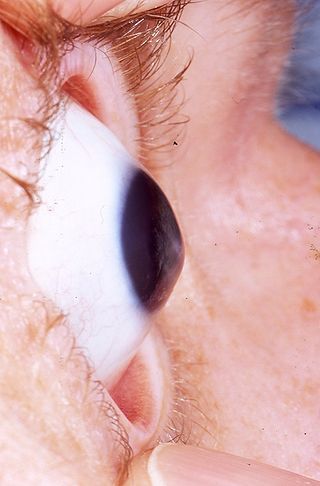
Keratoconus (from Greek: kerato, meaning horn and konos, meaning cone) is a progressive disorder of the cornea (the front of the eye), causing it to thin and change to a more conical shape than the more normal sphere of the cornea.
Keratoconus can cause substantial distortion of vision (multiple images, streaking, sensitive eyes, drop of vision are all often reported by patients). It is typically diagnosed in the patient’s adolescent years. It affects both eyes and visual deterioration can affect the patient’s ability to drive a car or read normal print. Patients typically have frequent changes in spectacle prescription before being diagnosed. Diagnosis usually requires careful clinical examination and corneal topography. Once the diagnosis is made, special contact lenses can improve vision. If left untreated, the cone is progressing and eventually the eye needs a corneal transplant for visual rehabilitation.
CROSS-LINKING (CXL)
The only treatment today that can stop progression is corneal collagen cross-linking. It involves a one-time application of riboflavin (vitamin B2) solution to the eye, which is then activated by illumination with ultraviolet light for approximately 30 minutes.The riboflavin causes new bonds to form across adjacent collagen fibers inside the cornea, which recovers and preserves the cornea’s mechanical strength. The corneal epithelial layer (surface layer) is generally removed to increase penetration of the riboflavin into the stroma, but it heals usually within 4 days.
Sometimes CXL can be combined with excimer laser ablation (down to 50 microns) to improve vision. Dr George Kampougeris has extensive experience with both techniques and would be happy to discuss available options in your case.
G. Kampougeris, MD, MRCSEd (Ophth), PhD Consultant Ophthalmic Surgeon, x Consultant, Moorfields Eye Hospital NHS Trust
Private Practice Office
CHALANDRI INSTITUTE OF OPHTHALMOLOGY Katsoulieri 4 st., 1st floor at 'FLIA' Mall Chalandri, Athens, Greece